A dental X-ray is an image of your oral cavity that your orthodontist uses to evaluate your dental hygiene. These X-rays, which employ low doses of radiation, are used to acquire pictures of the interior of your teeth and gums. This can assist your dentist in identifying issues such as cavities, dental decay, and affected teeth.
It’s natural for parents to worry about their child’s safety, especially during a treatment like an x-ray. The x-rays are quick, and the Tooth Fairy Dental Clinic ensures that your child is relaxed and secure.
Dental X-rays are classified into two types: intraoral (meaning the X-ray film is within the mouth) and extraoral (meaning the X-ray film is on the mouth’s outer side)
- The most basic pattern of dental X-rays done is an intraoral X-ray. These X-rays give a great deal of information and allow your dentist to discover cavities, examine the condition of the tooth root and bone around the tooth, assess the status of growing teeth, and monitor the overall health of your teeth and jawbone.
- Extraoral X-rays reveal teeth, although the jaw and skull are the primary focus. Because these X-rays lack the resolution present in intraoral X-rays, they are not used to detect cavities or diagnose abnormalities with individual teeth.
Extraoral X-rays are operated to search for affected teeth, evaluate jaw growth and development concerning the teeth, and detect possible issues between teeth and jaws and the temporomandibular joint or other bone compositions of the face.
Intraoral X-Rays, the Various Types
There are various forms of intraoral X-rays, each of which reveals a distinct element of the teeth.
- Bite-wing X-rays provide information on the upper and lower teeth in a single area of the mouth. Every bitewing represents a tooth right from the crown, to the underlying bone level. Bite-wing X-rays are employed to identify tooth decay and bone density changes brought forth by gum disease. They can also be used to assess the fit of a crown (or cast repair) and the marginal integrity of fillings.
- Periapical X-rays show the complete tooth, from the crown completely down to the end of the root, as well as the location of the tooth in the jaw. Each periapical X-ray displays the whole tooth size and includes all of the teeth in one section of the upper or lower jaw. These particular types of x-rays are used to find out certain inconsistencies in the bone composition as well as the root structure.
- Occlusal X-rays are more detailed and reveal complete tooth growth and positioning. Every X-ray image depicts the complete range of teeth in the lower or upper jaw.
Extraoral X-Rays, the Various Types
Extraoral X-rays can be taken in a variety of ways, depending on your dentist’s preference.
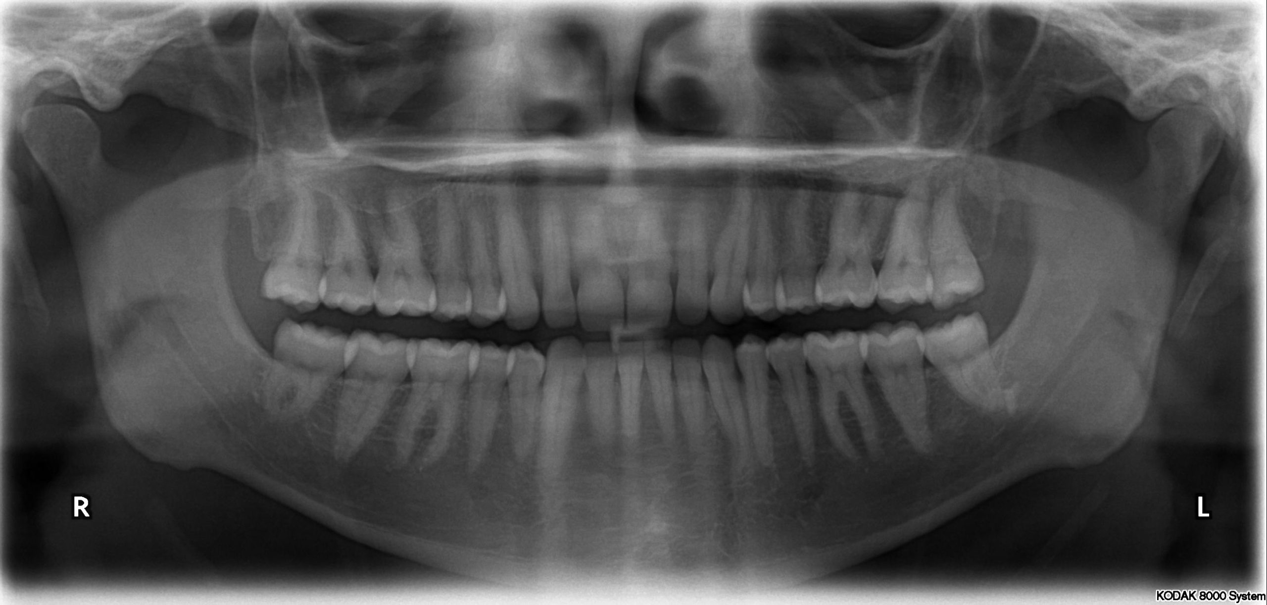
- Panoramic X-rays show the complete mouth region on a single X-ray, including all of the teeth in both the upper and lower jaws. This form of X-ray is beneficial for detecting the location of completely emerged as well as emerging teeth, identifying impacted teeth, and aiding in tumor identification.
- Tomograms display a specific layer or “slice” of the mouth while fading out the rest. This sort of X-ray is beneficial for investigating components that are challenging to see properly, such as when other structures are in near vicinity to the component to be examined.
- The full side of the head is shown in Cephalometric projections. This sort of X-ray is beneficial for assessing the teeth concerning the individual’s jaw and profile. This sort of X-ray is used by orthodontists to generate treatment plans.
- Sialography is the examination of the salivary glands after a dye has been injected. A radiopaque contrast chemical is administered into the salivary glands to allow the organ to be viewed on the X-ray imaging. Dentists may request this sort of test to examine for abnormalities with the salivary glands, such as blockages or Sjogren’s syndrome.
- CT scanning, or computed tomography, creates a three-dimensional picture of the body’s inner architecture. This X-ray, which is done at a hospital rather than a dentist’s office, is used to detect issues with the bones of the face, such as tumors or fractures.